Back pain, sciatica and lumbar radiculopathy are among the most common reasons patients seek medical advice and imaging. Yet one of the biggest challenges facing healthcare professionals is knowing when symptoms can continue to be managed conservatively and when referral for specialist assessment may be appropriate.
At the Oryon Academy Spinal Symposium 2026, consultant spinal surgeon Michael Hess explored this important topic in his presentation, Lumbar Radiculopathy – When Escalation Is Appropriate. Drawing on decades of experience and a series of real-world patient cases, he shared practical insights into recognising red flags, interpreting MRI findings and identifying when specialist intervention may be needed.
Watch the Full Presentation
This article summarises the key learning points from Michael Hess’s presentation. To watch the full session, including case studies, MRI examples and discussion around surgical decision-making, click below.
▶ Watch the full presentation: HERE
What Is Lumbar Radiculopathy?
Lumbar radiculopathy occurs when one or more nerves in the lower spine become compressed, irritated or inflamed. Patients often experience symptoms that radiate from the lower back into the buttock, leg or foot.
The most common cause is a lumbar disc herniation, often referred to as a slipped disc, which accounts for the majority of cases in adults under the age of 60. Other causes include spinal stenosis, foraminal stenosis, degenerative changes and synovial cysts.
Common symptoms include:
- Sciatica
- Shooting or burning leg pain
- Tingling or pins and needles
- Numbness
- Muscle weakness
- Reduced reflexes
- Difficulty walking
- Foot drop
Although these symptoms can be extremely distressing, the severity of pain alone does not always reflect the severity of the underlying condition.
Understanding the Difference Between Symptoms and Signs
One of the key themes of Michael Hess’s presentation was the importance of distinguishing between symptoms and clinical signs.
Patients often describe severe pain, but pain itself is a symptom. Clinicians must also look for objective neurological signs that indicate nerve root involvement.
These include:
- Sensory changes and numbness
- Muscle weakness
- Changes in reflexes
- Dermatomal sensory loss
- Myotomal weakness patterns
A thorough clinical examination remains essential when assessing patients with suspected lumbar radiculopathy. MRI findings should always be interpreted alongside the patient’s symptoms, neurological examination and functional limitations.
Sciatica and Disc Herniation: Does MRI Always Tell the Full Story?
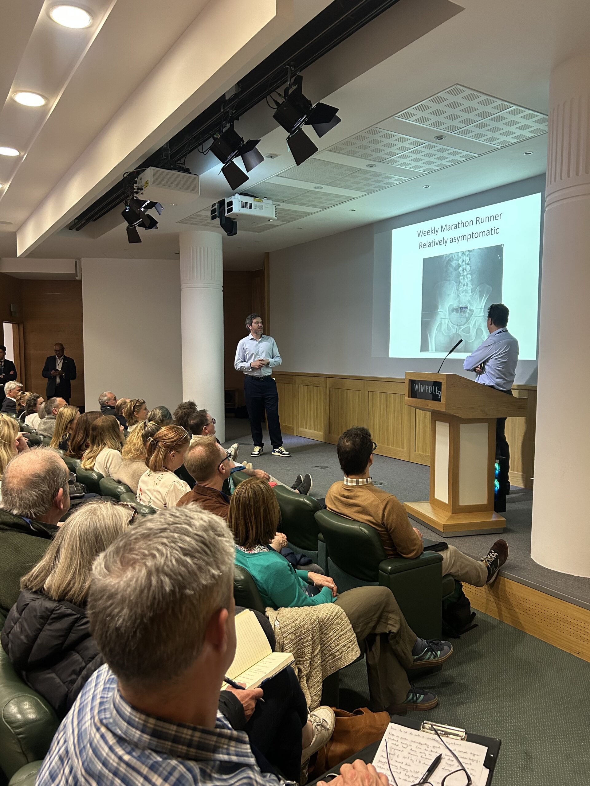
Many clinicians assume that larger disc herniations automatically cause more severe symptoms. However, Michael Hess demonstrated that this is often not the case.
Through a series of patient examples, he highlighted how MRI findings and symptom severity do not always correlate.
Some patients with significant disc herniations experience relatively mild symptoms and maintain good function, while others with less dramatic imaging findings may experience debilitating pain, difficulty walking or substantial neurological deficits.
The key message is that an MRI scan can help explain neurological signs, but it does not always explain the patient’s experience of pain.
This is why clinical assessment remains just as important as imaging when deciding whether escalation is appropriate.
Recognising Red Flags: Cauda Equina Syndrome
While many cases of sciatica and lumbar radiculopathy can initially be managed conservatively, clinicians should remain alert to symptoms that may indicate cauda equina syndrome.
Potential warning signs include:
- Saddle anaesthesia
- Urinary retention
- Bladder dysfunction
- Bowel dysfunction
- Sexual dysfunction
- Progressive neurological deficit
Cauda equina syndrome is a spinal emergency requiring urgent assessment and treatment.
Michael also emphasised the importance of maintaining a broad diagnostic perspective. Not every patient presenting with neurological symptoms has a straightforward spinal cause, and clinicians should remain open to alternative diagnoses when symptoms do not fit the expected pattern.
When Should Patients Be Referred?
One of the most valuable aspects of the presentation was the discussion around referral timing.
Many patients improve with conservative management, including physiotherapy, activity modification, medication and injection therapies. However, some patients may benefit from earlier specialist review.
Progressive Neurological Deficits
The presence of objective neurological weakness is often more significant than pain severity alone.
Examples include:
- Foot drop
- Quadriceps weakness
- Progressive muscle weakness
- Loss of function
- Significant sensory deficits
When neurological deficits are worsening, referral to a spinal specialist should not be delayed.
Severe Persistent Radicular Pain
Patients with severe leg pain that is significantly affecting quality of life and has not responded to appropriate conservative treatment may also warrant specialist assessment.
Persistent symptoms can affect mobility, sleep, work and mental wellbeing, and some patients may benefit from earlier intervention.
Correlation Between Symptoms and Imaging
Perhaps the most important principle discussed was ensuring that MRI findings genuinely explain the patient’s symptoms and neurological signs.
Referral decisions should not be based on imaging findings alone.
When symptoms, examination findings and MRI results align, treatment decisions become clearer. Conversely, caution is warranted when imaging abnormalities do not correlate with the patient’s clinical presentation.
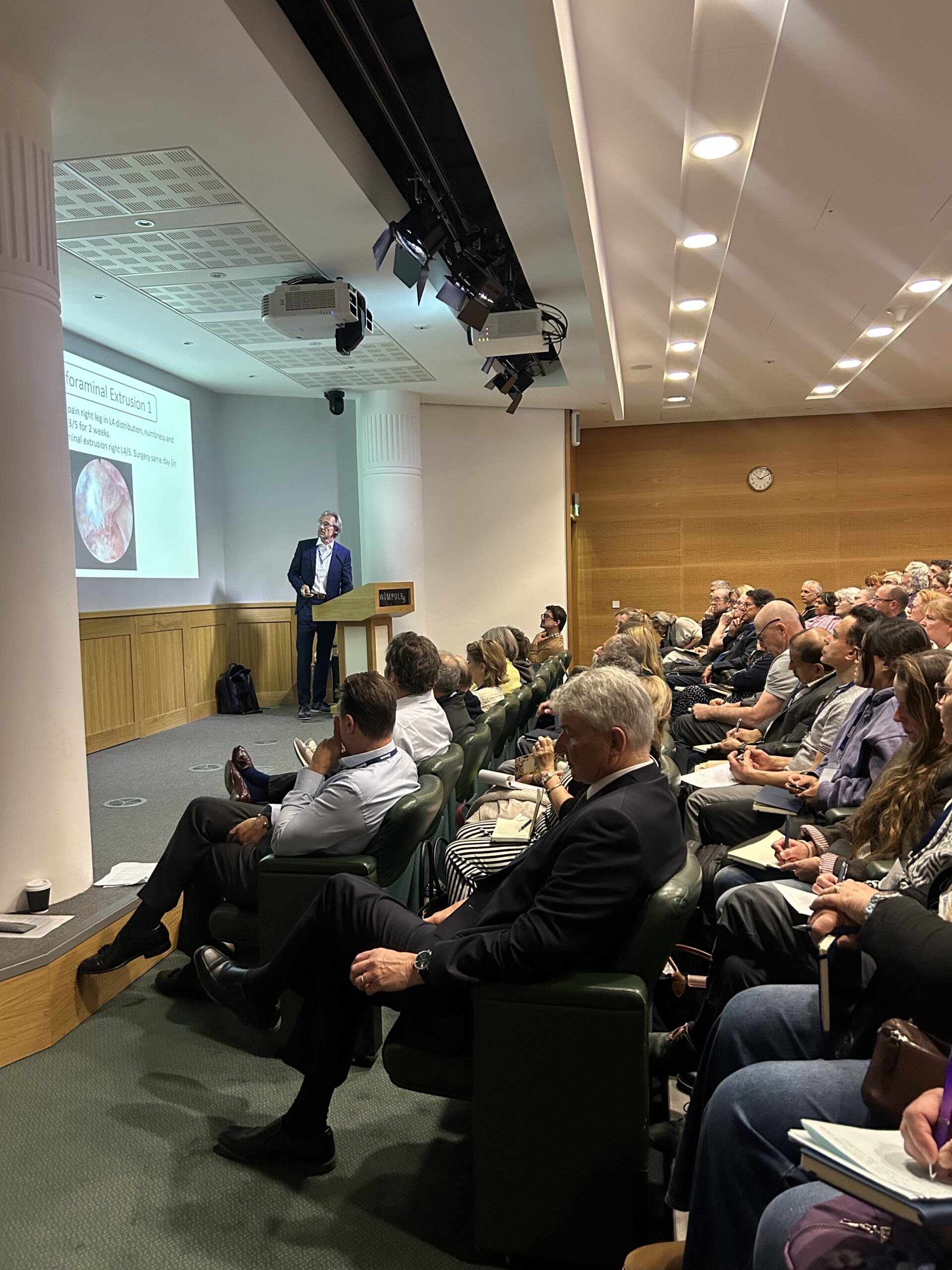
Foraminal Stenosis, Foot Drop and Other Causes of Nerve Compression
While lumbar disc herniation is the most common cause of lumbar radiculopathy, Michael Hess presented several cases involving other pathologies that can cause severe symptoms.
These included:
- Foraminal stenosis
- Intraforaminal disc extrusion
- Synovial cysts
- Recurrent disc herniation
Several patients presented with significant weakness, numbness and foot drop despite prolonged periods of conservative treatment.
These cases reinforced the importance of recognising when symptoms may reflect ongoing nerve compression that could require specialist intervention.
The Role of Endoscopic Spine Surgery
Michael Hess also discussed advances in minimally invasive spinal surgery, particularly endoscopic spinal procedures.
Endoscopic discectomy and decompression techniques are increasingly being used to treat selected patients with lumbar disc herniation, foraminal stenosis and nerve root compression.
Potential benefits may include:
- Smaller incisions
- Less tissue disruption
- Reduced post-operative pain
- Faster recovery
- Day-case treatment for many patients
- Shorter hospital stays
Several of the cases presented demonstrated significant improvements in leg pain, mobility and quality of life following endoscopic treatment.
As with all interventions, careful patient selection remains critical.
Key Takeaways for Referrers
Michael Hess concluded his presentation with several practical messages that are highly relevant for GPs, physiotherapists, osteopaths, chiropractors, sports medicine clinicians and spinal specialists.
Think Beyond the MRI
Imaging findings alone should not drive treatment decisions. Clinical examination and neurological assessment remain essential.
Assess Neurological Signs Carefully
Weakness, numbness and reflex changes often provide more useful information than pain severity alone.
Correlate Symptoms With Imaging
Patients benefit most when MRI findings clearly explain their symptoms and examination findings.
Avoid Operating on MRI Findings Alone
Not every abnormality seen on MRI is responsible for the patient’s symptoms.
Don’t Let Patients Suffer Unnecessarily
When symptoms are severe, persistent or associated with neurological deficits, timely referral can make a significant difference to patient outcomes.
Remember That Escalation Is Individual
Outside of emergency conditions such as cauda equina syndrome, referral and treatment decisions should always be tailored to the individual patient.
Watch the Full Presentation
Michael Hess’s presentation offers valuable insights into the diagnosis and management of lumbar radiculopathy, sciatica and lumbar disc herniation, supported by real patient cases and practical clinical examples.
▶ Watch the full presentation: HERE
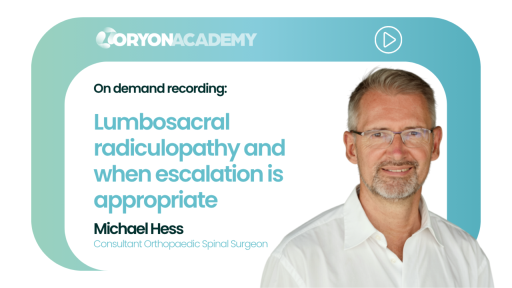
About Michael Hess
Michael Hess is an internationally recognised consultant spinal surgeon specialising in the diagnosis and treatment of lumbar disc herniation, sciatica, spinal stenosis, nerve root compression and complex spinal conditions.
His expertise includes minimally invasive and endoscopic spinal surgery, helping patients access effective treatment while minimising recovery times where appropriate.
Learn More
- ▶ Watch the full presentation: HERE
- View Michael Hess’s practitioner profile: https://oryon.co.uk/practitioners/mr-g-michael-hess
- Explore Oryon’s spinal imaging services
- Refer a patient to Oryon Imaging
Frequently Asked Questions
What is lumbar radiculopathy?
Lumbar radiculopathy occurs when a nerve root in the lower spine becomes compressed or irritated, causing symptoms such as sciatica, numbness, weakness and leg pain.
What is the difference between sciatica and lumbar radiculopathy?
Sciatica refers to pain travelling along the sciatic nerve pathway. Lumbar radiculopathy is the underlying neurological condition that often causes sciatica symptoms.
Can a lumbar disc herniation heal without surgery?
Many lumbar disc herniations improve with time and conservative management. Treatment decisions depend on symptom severity, neurological findings and patient-specific factors.
When should a patient with sciatica be referred to a spinal specialist?
Referral may be appropriate when symptoms are severe, persistent, worsening or associated with neurological deficits such as weakness, numbness or foot drop.
What does an MRI show in lumbar radiculopathy?
A spinal MRI can identify disc herniations, nerve root compression, spinal stenosis, foraminal stenosis and other structural causes of lumbar radiculopathy, helping clinicians correlate imaging findings with symptoms and examination findings.