Get in touch 020 7042 1888 | imaging@oryon.co.uk


The Future Of Radiology In Healthcare
With Technological Advancements, How Will It Evolve?
The Future Of Radiology In Healthcare
Posted on Tue Nov 9, 2021
In radiology, imaging technology plays an essential role in the diagnosis and treatment of diseases.
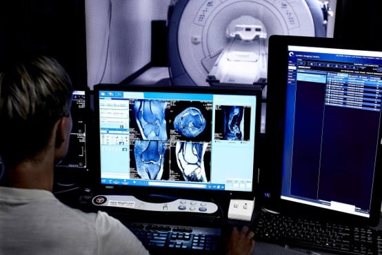
The images are taken by medical imaging professionals known as Radiographers using specialist imaging equipment. These images are then used by medical doctors known as Radiologists to interpret images of the body’s organs to diagnose disease. Based on their findings, radiologists will then compose a detailed report which are then provided to referring clinicians who can range from surgeons, GPs to other Allied Health Professionals.
Why is radiology important in medicine, and how important is it in making a diagnosis?
Several imaging modalities are available in radiology, each have their own physical principle, which varies in complexity and how crucial they are to medical care.
Some of these modalities include:
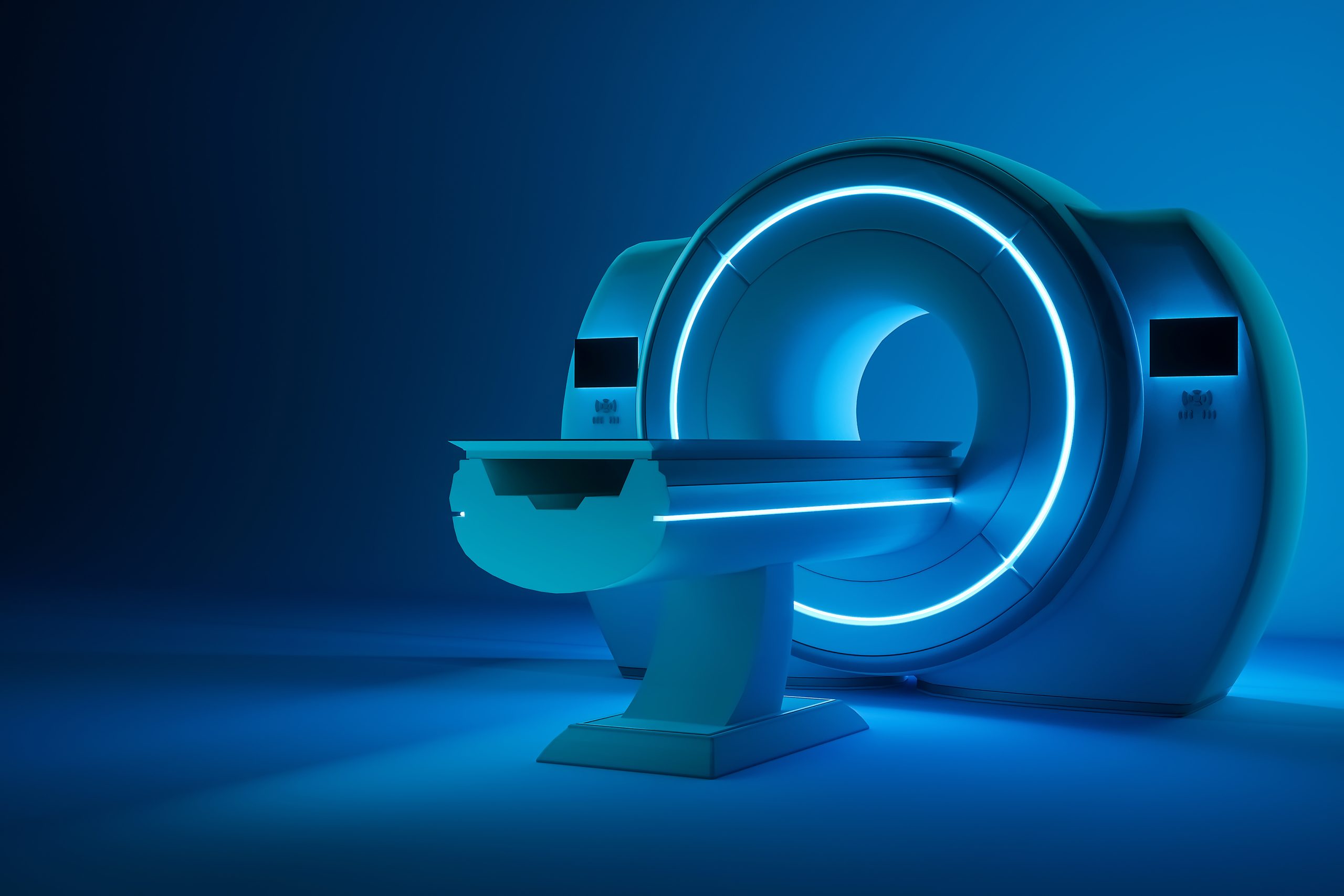
- MRI: A powerful device that creates images using magnetic fields
- CT Scan: This device uses X-rays to get detailed images of the body noninvasively
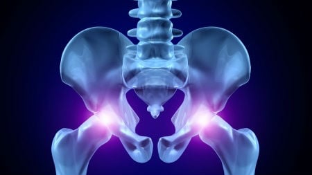
- X-rays: A device that uses ionising radiation to generate images of tissues and structures inside the body for instance, having a foot X-ray.
- Ultrasound: A small device known as a probe generates high-frequency sound waves to identify changes in the body’s internal organs.
- MRI: DEXA (Bone densitometry): This device uses a minimal dosage of ionising radiation (X-rays) to produce images of the body internally.
- Fluoroscopy: An imaging technique that uses X-rays to acquire images that are moving in real-time.
These devices are all key diagnostic tools that can help monitor treatments and are also essential in helping to foresee specific outcomes.
Radiology continues to be a crucial part of medical diagnostics and disease management. It allows physicians to gain detailed information about the bodies structure or underlying illness-related changes. Through having the capacity to diagnose during the beginning phases, diseases and injuries can be attended to, and patients might be saved. Without radiology, this may not be at all possible.
Click here to read more about the use of diagnostic imaging and the effect that increased demand has had on the public and private sector, with our Consultant Radiologist, Dr Qaiser Malik.
How is Artificial Intelligence being used in radiology?
Radiology is an integral part of medical care and one of the most powerful diagnostic and treatment tools available. So, let’s take a deeper look into how artificial intelligence is being used in radiology.
Artificial intelligence is a technology that uses computerised algorithms – which are step-by-step procedures for calculations, data processing and automated reasoning. Information is gathered over time to form algorithms that will achieve the best possible outcomes for an AI- this is an essential part of machine learning. This information is then followed to accomplish a goal or solve a problem.
Regarding radiology, the computer code written based on the algorithm will take the medical image as data input and return an answer to help the radiologist with their analysis.
What are the benefits of AI in radiology?
Research has shown that there has been greater accuracy and specificity using a computer (AI) in diagnostic medical imaging- they can pick up abnormalities. They are helpful in picking up repetitive tasks. These tasks typically involve a lot of data that radiologists would generally have to do, but they can now spend their time focusing on other important areas. It also helps to find subtle lesions. This is especially useful when the radiologist may be tired or distracted by other things. An additional benefit to having an AI is that they can provide a second opinion by analysing the physician’s diagnostic results.
How might AI help the radiologist help patients?
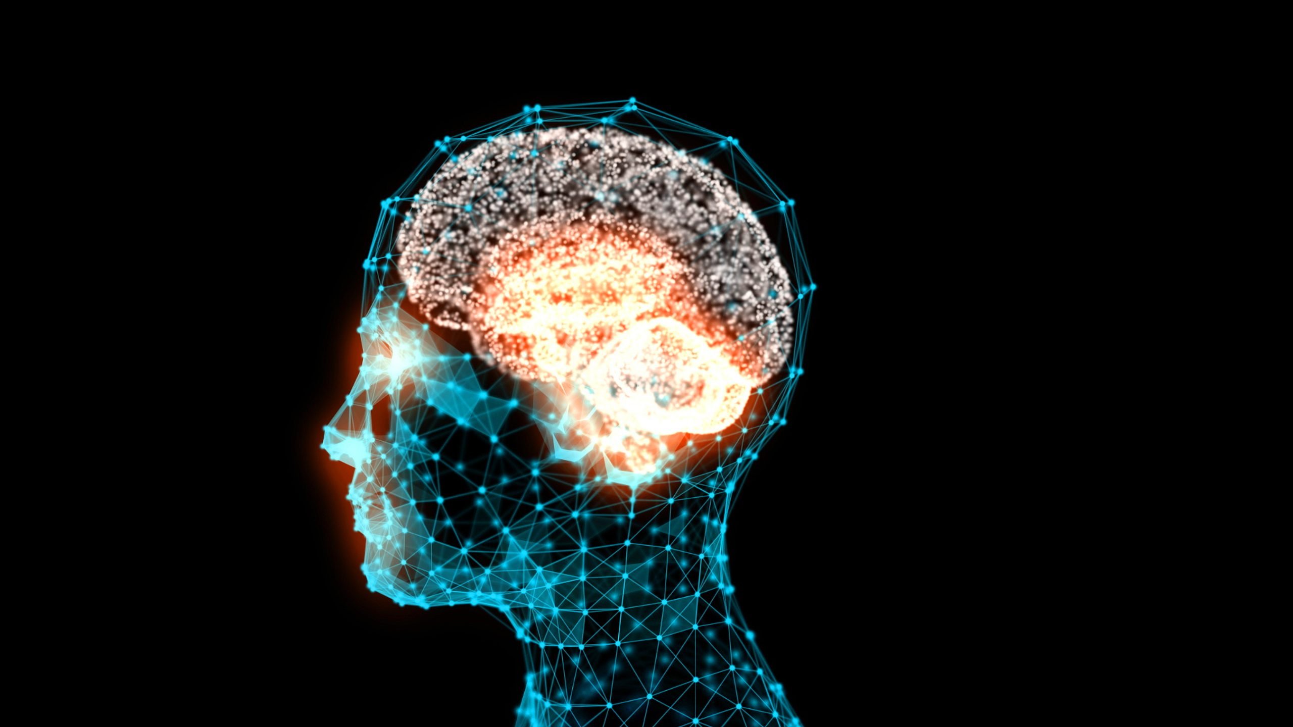
Artificial intelligence using Google’s DeepMind algorithm can detect breast cancer more accurately than a real doctor (Nature –www.nature.com). These findings came through research from Imperial College London and Google Health, where they had “trained” a computer to spot abnormalities on X-ray images of up to 29,000 women.
Increasingly, medical imaging is becoming part of the diagnostic chain. So, in effect, it is important that the end goal of the AI be the same as the radiologist; it must be beneficial to the patient’s wellbeing.
Conclusion
Timing is very important for health and giving a diagnostic earlier on could make a great amount of difference to saving a patient’s life. Computers can process large amounts of data, allowing them to perform analytical tasks beyond human capabilities.
The question remains could the future of AI development in radiology be the factor that saves numerous lives?
Share this article
Most Recent
What To Expect From a Prostate MRI
Posted on Fri Oct 3, 2025
Interview with Our Referral Relationships Manager
Posted on Fri Oct 3, 2025
Posted on Tue Sep 16, 2025