Mr Bal Dhinsa, Foot & Ankle Surgeon, examines 6 of the most common ankle injuries incurred whilst playing football.
Ankle injuries are common in professional and amateur level football, with the majority being lateral ligament injuries. The foot and ankle have to withstand great demands placed upon it during normal daily activities, however playing football increases the forces placed upon it, with the additional impact of legal and illegal contact with opponents.
1. Lateral ligament injury
This typically occurs when the foot is rolled inwards, either from a mis-step or following a tackle. The injury tends to be worse if the foot is planter-flexed at the time of impact. Often associated with a sudden change of direction, there are grades of sprain severity, up to complete rupture.
Short-term management
If there is concern regarding a possible fracture it is important to seek attention from a medical professional to assess and arrange investigations (such as plain radiographs).
With soft tissue injuries, initial resting of the limb, application of ice, high elevation (above the heart level) and compression should be instigated. Getting the ankle moving early with a rehabilitation programme that takes into account the degree of swelling and pain present at each stage is important for ligament healing.
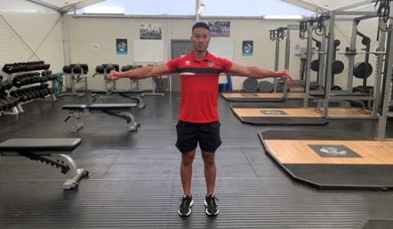
Proprioceptive training is key to the rehabilitation process. This is specific balance exercises used to improve the awareness of the ankle joint position. These exercises will help prevent re-occurrence of any injuries. There are some exercises you can do yourself, but I would advocate seeking the advice of a physiotherapist prior to commencing.
As the ankle feels more stable, pain and swelling reduces, rehabilitation can progress to more functional exercises.
Avoidance of injuries
Unfortunately, if you have had an ankle injury you will be at greater risk of further injuries to the ankle. This is often the case if players have poor rehabilitation and return to the pitch too quickly.
The proprioceptive exercises mentioned above will help to strengthen/stabilise the ankle joint and help reduce the risk of further injury. Other factors that can be of benefit include losing excess weight (thus reducing load on the ankle) and assessing for abnormal biomechanics of the foot and ankle that can be potentially managed with physiotherapy or orthotics.
When to seek medical advice
In the initial stage, if there is a concern about possible fracture or associated injuries it is sensible to seek medical advice to rule this out. With the professional athlete, a thorough consultation is required to discuss the role of acute operative reconstruction and functional/proprioceptive rehabilitation.
If the ankle remains unstable despite a thorough rehabilitation process, or in the case of reoccurrence of injury, a medical opinion should be sought for consideration of operative measures. The instability is often the result of the injured ligaments not healing or remodelling with an elongated length. Subsequently, the ankle has a range of motion greater than normal limits with further strain on the capsule and soft tissues affecting the proprioceptive feedback.
2. Osteochondral lesions
The cartilage lining the ankle joint is frequently damaged with sprains, as the joint surfaces impact on each other. There is initially a localised bruise, followed by cartilage softening and then a small crack in the surface can develop. If this progresses a cyst may form in the bone below the cartilage surface, forming an osteochondral lesion and sometimes a piece of cartilage may become loose.
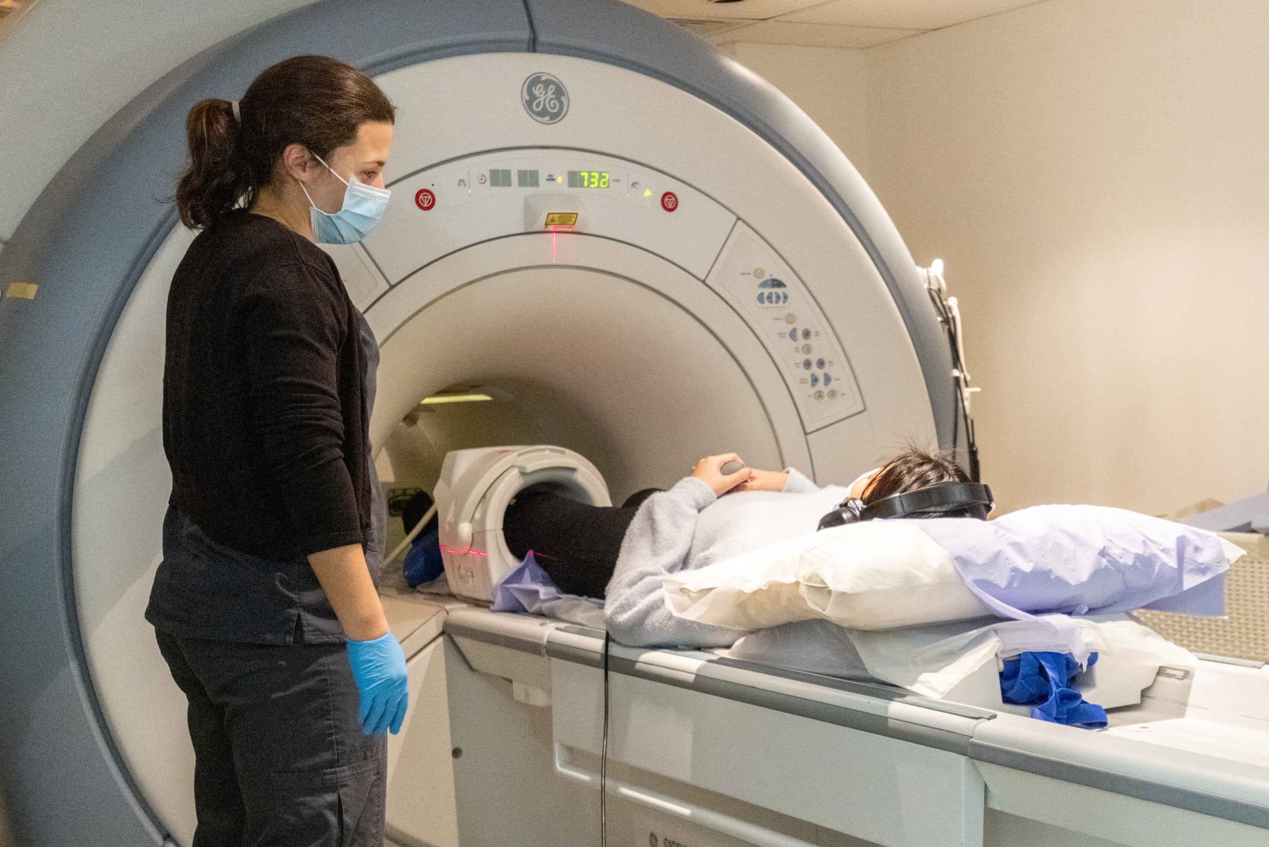
Pain is typically felt in the joint and becomes worse with activities. With careful examination the specific location can be isolated to either the medial, central or lateral aspects of the ankle. Patients may also have stiffness, locking or catching as a result of the injury and this may need addressing as well. If suspected, a magnetic resonance imaging (MRI) scan is recommended for diagnosis, as well as assessment of location and size of lesion.
Management
Initial management for all lesions should be protection of the joint (ankle brace or walking boot), rest (crutches), compression (sometimes a compression bandage is of help) and elevation. Analgesics and non-steroidal anti-inflammatory medication may also be used. Corticosteroid intra-articular injections may also be considered.
When to refer onwards
Most osteochondral lesions will not heal due to the poor blood supply. With persistent pain and symptoms despite non-operative measures, arthroscopic (‘keyhole’) surgery can be considered for small lesions to stimulate the bone marrow. For the larger lesions, greater than 15 mm, than open surgery through a small incision at the front of the ankle (arthrotomy) may be required to fill the defect with graft.
3. Ankle impingement & soft tissue impingement
This typically occurs in the anterolateral aspect of the ankle and results from entrapment of inflamed and/or chronically damaged soft tissue. The player complains of limited ankle dorsiflexion motion and swelling after training/matches. MRI can be of use, as well as a diagnostic/therapeutic injection of local anaesthetic and corticosteroid.
Management
Mainstay of initial management is physiotherapy and deep tissue massage, as well as following the RICE protocol during acute episodes. If there is no improvement with these measures and injections, it may be necessary to undergo surgical intervention. This would be an arthroscopic assessment and debridement. Unfortunately, reoccurrence of impingement after surgical intervention is possible.
Bony impingement
This is impingement resulting from osteophytes (bone spurs), usually in the anteromedial aspect of the ankle, leading to reduced dorsiflexion of the ankle. The cause is not fully understood but is thought to be from repetitive microtrauma to the ankle. Presentation is similar to soft tissue impingement, however typically tenderness to palpation is felt on the anteromedial aspect. Plain radiographs can be of help, and augmentation with an ankle MRI scan helps to look for associated injuries.
When to refer
Initial management is the same as for soft tissue impingement, however it is often less effective given the presence of a mechanical block to motion. Operative management is arthroscopic debridement to improve range of motion, however it is not uncommon to have to repeat the procedure in the future for reoccurrence.
4. Plantar Fasciitis
The high plantar pressures seen in training and matches, particularly on artificial surfaces, predisposes players to plantar fasciitis. A sudden increase in activity places the players at increased risk.
Management
The majority of players can be managed with resting, a rehabilitation programme including plantar fascia specific stretching exercises, and the use of orthotics. Extra-corporeal shockwave therapy may also be used to complement the physiotherapy. If these measures fail to improve symptoms, dry needling and corticosteroid injections may be indicated.
When to refer
If all the above measures fail, and there is calf tightness present, a surgical procedure to ease the tightness maybe required. A open plantar fascia release and spur removal is not recommended, however if there is nerve entrapment associated with it, this may need decompressing.
How to avoid
Daily stretching, as well as a dedicated warm-up regime, is essential to stretch the calf and foot musculature in preparation for increased activity.
5. Tendoachilles tendinopathy
With overuse or repetitive strain, the tendoachilles can be injured and even develop a tear. These injuries heal with scar tissue leading to localised swelling of the tendon. This can occur at either the site of Achilles insertion into the calcaneum (insertional tendinopathy) or within its mid-substance (non-insertional Achilles tendinopathy).
The compliance with a comprehensive rehabilitation programme is essential, as well as an understanding between the team and player about the need to not rush to return to play until fully recovered. Unfortunately, this year there have been examples in American sport teams of players suffering rupture of the tendoachilles following an early return to sports after a ‘calf strain’.
Typically, there is pain around the injured area with swelling, often worse in the morning before stretching and after activity. There may be thickening seen over the area of inflammation and this can make wearing footwear uncomfortable.
Management
Initial management includes activity modification, incorporating rest periods, modification of footwear with heel lifts and stretching exercises. Analgesics and non-steroidal anti-inflammatory medication may also be used. Early physiotherapy will be instigated to help with the recovery.
Next steps in management could be an injection of high-volume fluid around the tendon under ultrasound guidance. This helps to free the paratenon sheath from the tendon, which can become inflamed and adherent to each other. An alternative is extracorporeal shock wave to help break down the scarring that is often present with inflammation and allows the stretching exercises to be carried out effectively.
When to refer
With persistent pain and swelling surgical intervention can be considered. For the non-insertional tendinopathy, the unhealthy tendon is debrided and repaired. However, for insertional tendinopathy the Achilles tendon needs to be lifted from its insertion, debrided and the Haglund’s bump (calcaneal bony prominence) resected prior to reattaching the tendon back to the calcaneum.
How to avoid
Daily stretching, as well as a dedicated warm-up regime, is essential to stretch the calf and foot musculature in preparation for increased activity.
6. Fractures
Fractures of the ankle in football players usually involve high-energy mechanisms with significant displacement making acute surgical intervention necessary. Rarely, a fracture can occur at low-velocity without direct impact, which results in a minimally displaced fracture which can be managed with immobilisation for 6-8 weeks. Once there is evidence of clinical and radiological healing rehabilitation can be commenced.
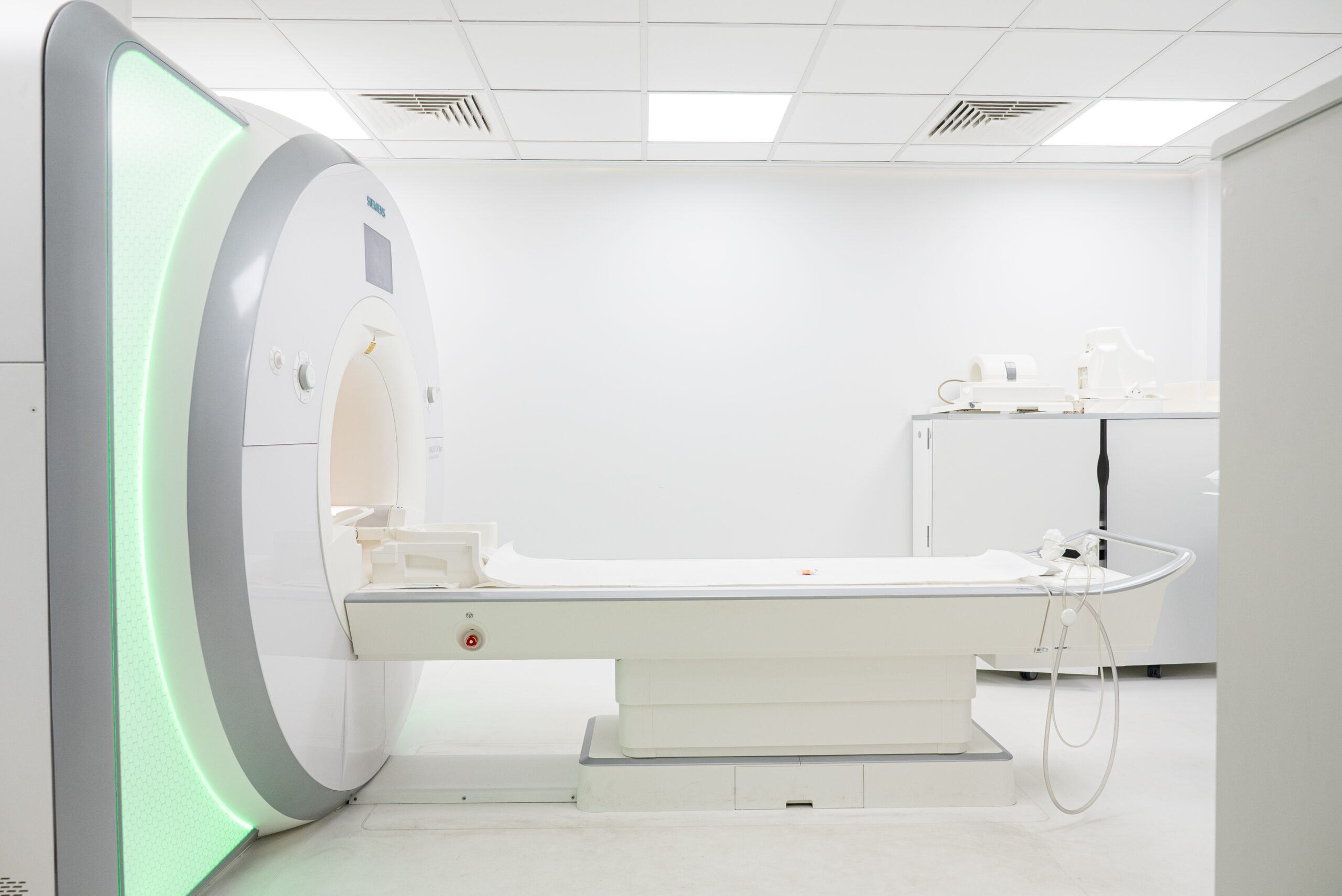
MRI of the ankle, showing an ankle fracture
Metatarsal fractures are frequently seen in football players, especially with the use of modern boots with less protection afforded to the foot in order to make them lighter. They may also occur as a stress fracture, especially if activity levels have suddenly increased. These fractures are often minimally displaced and can be managed with immobilisation for the initial 4-6 weeks to help with pain control, followed by physiotherapy. If the fracture is significantly displaced or has failed to heal after non-operative measures, surgical reduction and fixation maybe required. Unfortunately, any metalwork utilised in the fixation may need removal after fracture healing as athletes often find this uncomfortable.
Oryon Imaging
Oryon Imaging offers affordable private MRI scans, ultrasound scans, X-rays and DEXA scans in London.
About the author
Mr Bal Dhinsa is a Consultant Orthopaedic Surgeon, specialising in the foot and ankle. Visit his website here.